

 A team led by Columbia University researchers in New York established a new vascular technology for the diagnosis of peripheral arterial disease (PAD), a medical condition especially important in diabetic patients. The study was published in the European Journal of Vascular and Endovascular Surgery and is entitled “Detection of Peripheral Arterial Disease Within the Foot Using Vascular Optical Tomographic Imaging: A Clinical Pilot Study.”
PAD is a common vascular condition characterized by narrowed arteries due to the accumulation of plaques (composed of fat, cholesterol, fibrous tissue, among others) in a process known as atherosclerosis, which can cause reduced blood flow to the brain, organs and limbs, especially the legs. It is estimated that in the United States, approximately 8 to 12 million people suffer from this condition. PAD is known to increase the risk for stroke and heart attack by up to six times.
PAD is also considered one of the most serious complications in patients with diabetes, being responsible for over 71,000 leg amputations every year in this population, which corresponds to around 60% of all lower extremity amputations in the United States. As the number of diabetics is expected to increase, and being PAD seriously under-diagnosed due to the lack of a reliable screening method, an accurate and early diagnosis tool for this condition is therefore crucial to avoid further devastating health consequences.
Dr. Andreas Hielscher at Columbia University is currently developing a new, non-invasive diagnostic technology for PAD to aid in patient monitoring — a dynamic optical tomographic imaging system. This new technology is based on near-infrared light that allows the detection of hemoglobin concentration (the molecule respo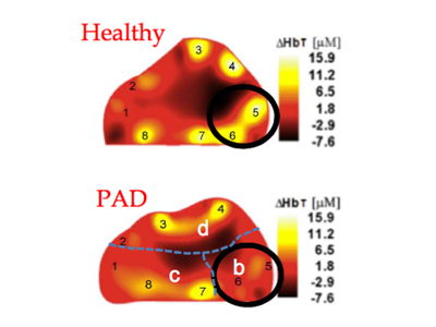 nsible for oxygen and carbon dioxide transport in red blood cells) in the tissues and to assess blood circulation in patients’ feet and hands. The intensity of the transmitted light is used to create spatially resolved maps of blood oxy- and deoxy-hemoglobin.
“We believe our new vascular optical tomographic imaging (VOTI) system will revolutionize the way we diagnose and monitor peripheral arterial disease,” said the study’s senior author Dr. Hielscher in a news release.
In this clinical pilot study, the team tested a prototype system of VOTI in 20 healthy controls and 20 patients with PAD (10 non-diabetic and 10 diabetic) for the diagnosis of the condition in the lower extremities.
Researchers found that the VOTI system was able to diagnose PAD and assess the blood circulation in various locations within the foot, allowing an evaluation of the arterial disease severity. This was also true for diabetic patients, who have calcified arteries and are therefore more difficult to assess and diagnose PAD.
“We’ve successfully used the VOTI to detect PAD in the lower extremities,” said the study’s first author Dr. Michael Khalil. “Unlike other methods, our VOTI technology can provide maps of oxy-, deoxy-, and total hemoglobin concentration throughout the patient’s foot and identify problematic regions that require intervention.”
The next goal of the team is to test the VOTI technology in larger clinical studies to assess vascular disease, especially in patients with diabetes. Another potential application for this non-invasive technology is the monitoring of wound healing.
Together with the New York City Partnership (NYCP) fund, Dr. Hielscher will commercialize certain elements of the VOTI technology and will develop a commercially viable device to be employed intra-operatively and guide interventions by vascular surgeons. The team believes that such a commercial device might be available within two years.
